Multimodal imaging relies on optimal integration of complementary technologies. Magnetoencephalography (MEG) and electroencephalography (EEG) directly reflect neural changes with excellent temporal resolution. However, underlying neural substrate cannot be inferred unambiguously from MEG/EEG. In contrast, hemodynamic assessment of brain activity using functional magnetic resonance imaging (fMRI) is an excellent spatial mapping tool. However, its temporal resolution is limited as it reflects neural changes only indirectly via neurovascular coupling. Multimodal imaging draws on respective advantages of these complementary methods to determine where the neural changes are occurring during cognitive tasks, and to understand the temporal sequence ("when") of the involved neural components.
Click on the links below for more information about our various methods!


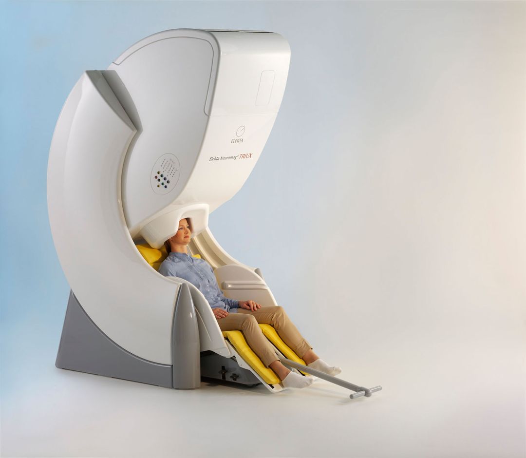
Magnetoencephalography (MEG) is a non-invasive imaging method that records magnetic fields produced by the brain with a millisecond precision. We use a 306-channel planar dc-SQUID* Neuromag Vectorview (Electa) system located at the UCSD Radiology Imaging Lab.
SQUID stands for "superconducting quantum interference device".
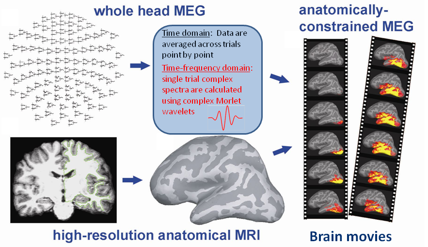
Our analysis employs an anatomically-constrained MEG (aMEG) approach which combines distributed source modeling of the MEG signal with each person's cortical surface reconstructed from high-resolution MRI scans. The resulting "brain movies" are spatio-temporal maps of activity estimates across time as a function of task conditions. The data are analyzed in both time- and time-frequency domains during cognitive tasks. Spontaneous oscillations recorded during wakeful rest are analyzed with the FFT-based algorithm in a wideband frequency range.
Check out our publication in the Journal of Visualized Experiments!
Disruption of Frontal Lobe Neural Synchrony During Cognitive Control by Alcohol Intoxication
Video Version: Text Version:
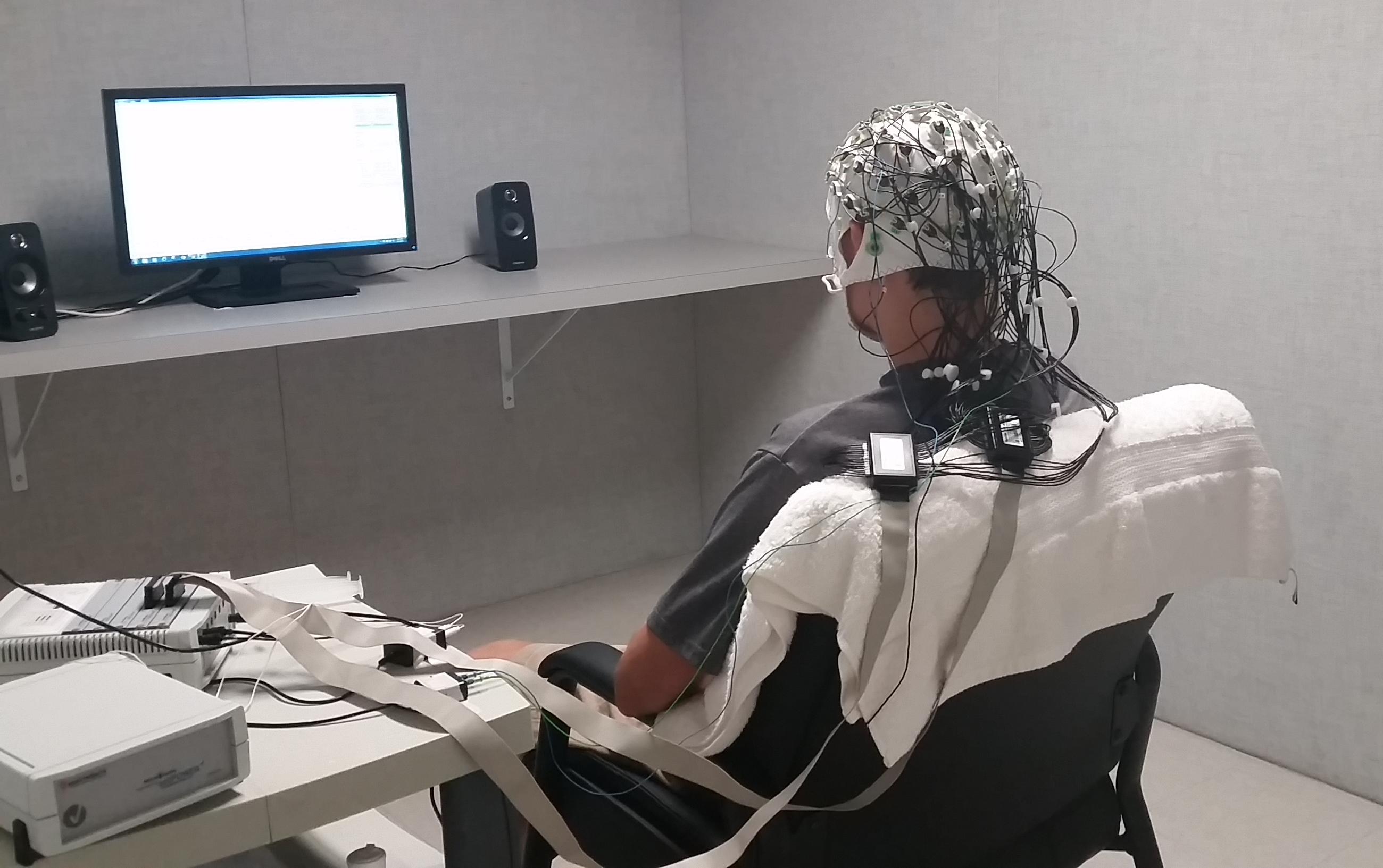
 Electroencephalography (EEG) measures electrical activity in real time and with a millisecond temporal resolution. We record EEG signals with a 64-channel actiCHamp DC Brain Vision System (Brain Products) and apply similar analysis methods as described above.
Our EEG studies take place at our lab at SDSU on Alvarado Road.
under construction...


