The overall goal of our lab is to examine spatio-temporal (i.e. “where and when”) characteristics of distributed neural circuits underlying cognitive and affective functions such as decision making, inhibitory control, language, and emotion perception. Our lab has many interests, including alcohol, Autism Spectrum Disorder, chronic pain, and others. We use multimodal functional imaging including magneto- and electroencephalography (MEG/EEG), functional and structural MRI, and psychophysiological measures of autonomic functions. The synergistic approach allows precise insight into on-line dynamics of these processes, with implications for individualized prevention strategies and pharmacogenetics.
In collaboration with Dr. Axel Mueller's lab we are investigating the neural correlates of language processing in individuals diagnosed with Austism Spectrum Disorder (ASD).
We have also collaborated with Dr. Thereasa Cronan's lab to investigate the effects of a treatment-based intervention program in individuals with fibromyalgia, as well as underlying biomarkers of symptoms.
If you are interested in participating in a study, please contact psych.STBIL@sdsu.edu (see the contact page for more info).
Click on the links below to learn more about the research we perform in the lab!
Our research focuses on the spatio-temporal characteristics underlying cognitive and executive functions in general, and as they pertain to alcohol effects. The synergistic approach based on integration of different brain imaging techniques is especially well-suited for obtaining highly precise insight into on-line dynamics of these processes, with an emphasis on location and sequence of alcohol-related impairments.
Our work, along with other evidence, shows that alcohol intoxication impairs decision making. When preferred or automatic responses must be overridden in lieu of a different response, alcohol interferes with this goal-directed behavior, and results in impaired self-control. Consequences of impaired self-control may be the inability to stop oneself from drinking. Our studies employ a battery of tasks which require participants to engage their cognitive control while we collect neuroimaging data, which is also integrated with personality measures and genetics.
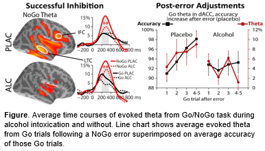
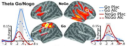
Group average maps of theta source power during successful response inhibition (NoGo) and execution (Go) and the right inferior frontal cortex (IFC). Alcohol attenuates theta to successful NoGo in the IFC.
Response Inhibition: Post-error Adjustments
Alcohol intoxication is associated with disinhibited, impulsive behavior. In this multimodal study, magnetoencephalography (MEG) was combined with structural MRI to examine the acute effects of alcohol on theta-band (4-7Hz) dynamics underlying inhibitory control, errors, and post-error optimization. Adult social drinkers participated in the Go/NoGo task during alcohol challenge.
Right-lateralized theta power elicited by successful response inhibition (NoGo) was attenuated by alcohol. Post-error engagement of cognitive control was reflected in increased dorsal ACC theta and accuracy, which was abolished by alcohol.
Average time courses of evoked theta from Go/NoGo task during alcohol intoxication and without. Line chart shows average evoked theta from Go trials following a NoGo error superimposed on average accuracy of these Go trials
Response inhibition and action cancellation engage a right-lateralized and includes the inferior frontal and supplementary motor cortices. In tasks such as Go/NoGo which require response inhibition, successfully withholding a prepotent response elicits theta power in a strongly right-lateralized network. Alcohol is also found to reduce theta in these regions during response inhibition.
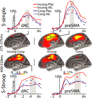
Average evoked theta timecourses are shown, sourced from the dAC and preSMA, also shown. Alcohol attenuates theta during response conflict (incong), especially during the more difficult task.
Decision conflict in ACC
Acute alcohol intoxication is known to attenuate the dorsal anterior cingulate (dAC), which is activated during decision/response conflict. We conducted a study which utilized simple and difficult versions of the Simon task to induce response conflict while participants were sober (PLAC) and during acute intoxication (ALC). Anatomically constrained MEG was used to measure task-related theta oscillations, which emerge from the dAC during response conflict.
It was found that alcohol intoxication generally reduced theta, however this effect was more pronounced during the more difficult version of the Simon task, and most prominently in the dAC. This is despite only a marginally lower reduction in accuracy for response conflict (incong) trials while intoxicated. Conflict related theta was also associated with weekly alcohol consumption, where greater theta reduction was seen among heavier drinkers.
Related Publications
Marinkovic, K., & Rosen, B. Q. "Theta oscillatory dynamics of inhibitory control, error processing, and post-error adjustments: Neural underpinnings and alcohol-induced dysregulation." Alcoholism: Clinical and Experimental Research (May 2022) PDF
Rosen, B. Q., Padovan N., and Marinkovic K. "Alcohol Hits You When It Is Hard: Intoxication, Task Difficulty, and Theta Brain Oscillations." Alcoholism-Clinical and Experimental Research 40, no. 4 (Apr 2016): 743-52.
Rosen, Burke Q., Ryan O'Hara, Sanja Kovacevic, Andrew Schulman, Nevena Padovan, and Ksenija Marinkovic. "Oscillatory Spatial Profile of Alcohol's Effects on the Resting State: Anatomically-Constrained Meg." Alcohol 48, no. 2 (Mar 2014): 89-97.
Kovacevic, Sanja, Sheeva Azma, Andrei Irimia, Jason Sherfey, Eric Halgren, and Ksenija Marinkovic. "Theta Oscillations Are Sensitive to Both Early and Late Conflict Processing Stages: Effects of Alcohol Intoxication." PLOS One 7, no. 8 (Aug 27 2012).
Marinkovic, K., Rickenbacher, E., Azma, S., Elinor A. "Acute Alcohol Intoxication Impairs Top-Down Regulation of Stroop Incongruity as Revealed by Blood Oxygen Level-Dependent Functional Magnetic Resonance Imaging." Human Brain Mapping 33, no. 2 (Feb 2012): 319-33.
Marinkovic, K., S. Azma, S. Kovacevic, E. Rickenbacher, J. Sherfey, A. Dale, and E. Halgren. "When the Executive Gets Drunk: Effects of Acute Intoxication on Cognitive Neurodynamics." Alcoholism-Clinical and Experimental Research 35, no. 6 (Jun 2011): 283A-83A.
Marinkovic, K., E. Rickenbacher, E. Artsy, and S. Azma. "Effects of Alcohol Intoxication on Saccadic Control: An fMRI Study." Alcoholism-Clinical and Experimental Research 33, no. 6 (Jun 2009): 88A-88A.
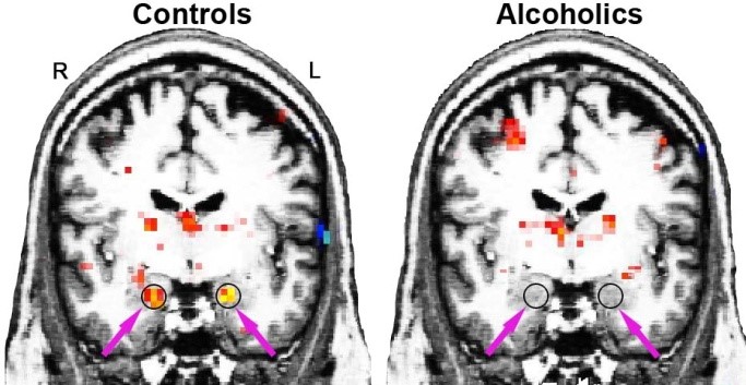
Circles and arrows show group averaged activations in right and left amygdalae. In sober participants with chronic alcoholism, activation in the amygdalae were blunted in response to emotionally expressive faces.
Studies of abstinent alcoholic individuals have indicated that excessive, chronic drinking can lead to brain damage on multiple levels, including atrophy and white-matter damage. The neuroimaging evidence from our research suggests that deficient activation of limbic structures inside the temporal lobes may underlie emotional abnormalities in abstinent long-term alcoholics including difficulties in accurate perception of emotional expressions. Misreading facial cues can escalate interpersonal conflict, creating liability for impaired social interaction and continued drinking. Future studies will determine to what degree these problems are the result of neural changes due to excessive alcohol consumption, or whether they precede the onset of alcoholism.
Related Publications
Marinkovic, Ksenija, Marlene Oscar-Berman, Trinity Urban, Cara E. O'Reilly, Julie A. Howard, Kayle Sawyer, and Gordon J. Harris. "Alcoholism and Dampened Temporal Limbic Activation to Emotional Faces." Alcoholism-Clinical and Experimental Research 33, no. 11 (Nov 2009): 1880-92.
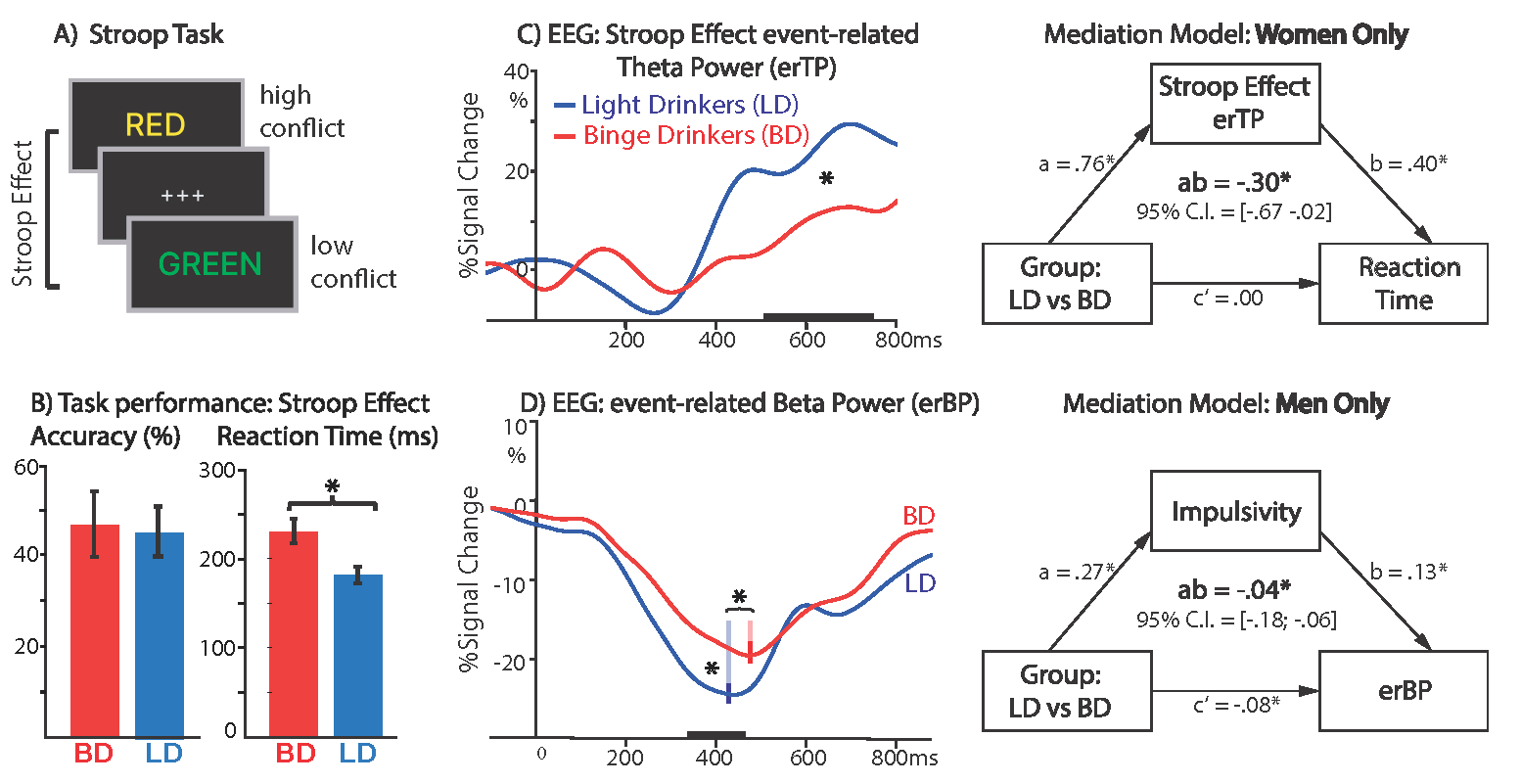
This EEG study examined cognitive and motor aspects of decision-making during a modified Stroop task (A) in young adult binge (BDs) and light drinkers (LDs). Besides compensatory response slowing to Stroop interference (B), BDs showed lower Stroop- induced theta power, which mediated the impact of BD on degraded task performance in BD women (C), indicative of impaired cognitive control. Conversely, impulsivity partially mediated an attenuated beta decrease during response preparation in BD men (D), suggesting sex-specific pathways of vulnerability in BDs.
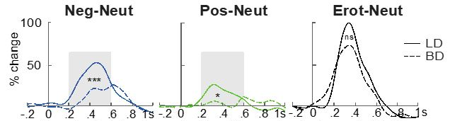
Emotion-induced event-related theta differences (emotional - neutral) within 200-600 ms over parietal electrode sites for light drinking (LD) and binge drinking (BD) groups. *p<.05, **p<.01, ***p<.001
Emotional processing
Binge drinking is selectively associated with attenuated event-related theta power elicited by emotional (vs. neutral) pictures across all scalp sites, although no group difference are observed on the subjective feelings towards the pictures. This finding indicates blunted widespread modulation of cortical processing and corticolimbic interactions which may underlie impaired emotional and social functions. This possible impairment is also consistent with compromised corticolimbic affection functions seen in alcohol addiction. Lowered theta power for emotional (vs. neutral) photos is positively correlated with the binge drinkers’ self-reported number of binge episodes during the past six months, suggesting these deficits were more pronounced for heavier drinkers.
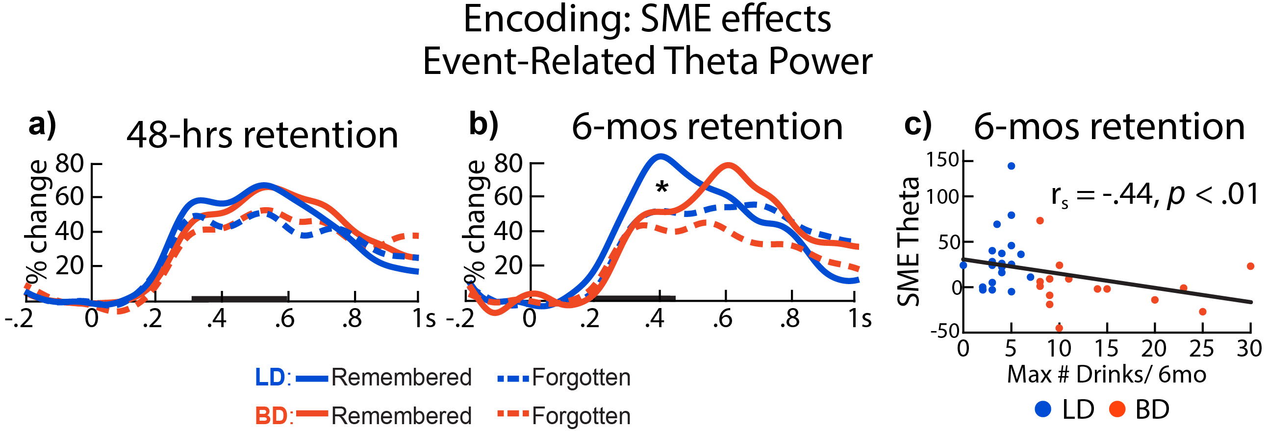
Event-related theta power during initial encoding associated with later remembered vs. forgotten pictures for LD and BD groups as a function of recognition (A) after 48 hs and (b) after 6 mos. Event-related theta power difference (remembered - forgotten after 6 mos) correlated negatively with self-reported maximum number o drinks consumed on a single occasion in the past 6 months *p<.05. Bolded bars on the x-axis mark the time windows of interest.
Memory Encoding
The same participants underwent a second retrieval session six months after the initial encoding session. Notably, the BD group exhibitied reduced theta oscillations and phase synchronization during the first presentation of images that they were able to remember six months later. Furthermore, the difference in evoked theta between remembered and forgotten images (SME) negatively correlated with the maximum number of drinks consumed in a day participants reported in the last six months. These findings suggest that memory encoding in binge drinkers may be characterized by inefficient network-level interactive engagement of brain areas responsible for memory formation, which may result from excitation/inhibition dysregulation as a function of increased drinking.
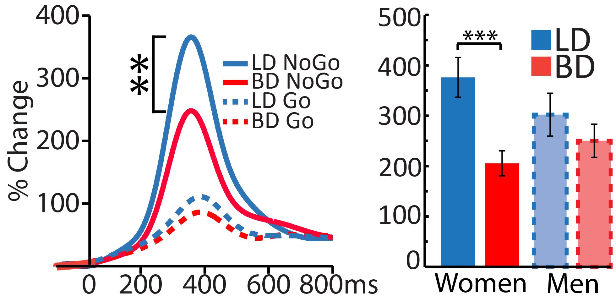
Group average time courses for event-related theta power. Looking at the peak of evoked theta, NoGo trials normally exhibit higher theta due to the inhibition of a prepotent response. However, BD participants had reduced NoGo theta power compared to LD. **p<.01.
Response Inhibition
In a Go/NoGo task which frequently requires the inhibiting of a prepotent motor response, NoGo (response inhibition) trials elicit much greater theta (4-7 Hz) activity than Go (response activation) trials. However, Binge Drinkers (BD) exhibit attenuated theta power, specifically on NoGo trials compared to Light Drinkers (LD). However, no group differences are seen in Go trials, suggesting that binge drinking selectively impairs response inhibition.
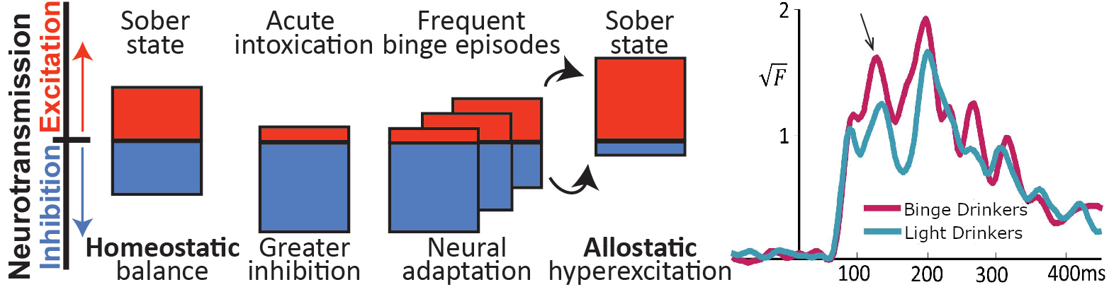
Schematic describing the allostasis model. Acute intoxication inceases inhibition, but the brain adapts by increasing excitation. After repeated binge drinking episodes, these adaptive changes lead to an overall increase om excitability even when sober. Timecourse depicts larger early visual response among sober binge drinkers, suggesting heightened excitability.
Allostasis
Acute alcohol intoxication descreases neural responsivity due to the pharamcological effects of alcohol. However, according to the allostasis model, chronic and repeated binge drinking can result in adaptive changes persisting beyond the acute drinking episode. In accordance with this model, the brain downregulates GABA-mediated inhibitory signaling and upregulates excitatory glutamatergic function, leading to increased neural excitability.
In support of this theory, our lab utilized a study which presented visual stimuli to sober light drinkers and binge drinkers. The findings revealed that the early visual response was attenuated in binge drinkers compared to light drinkers. This early response suggests heitghtened neural excitability, thereby corroborating the allostasis model in sober binge drinkers.
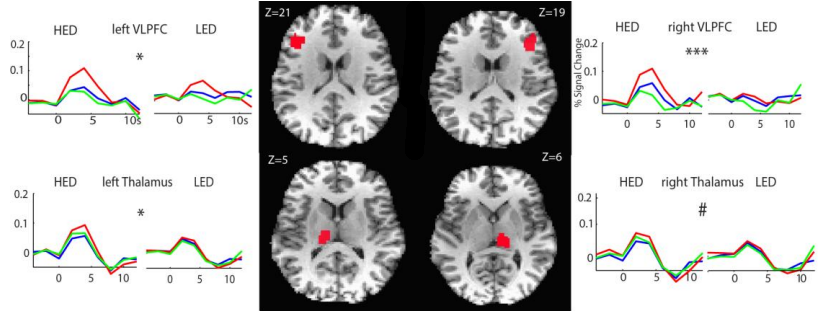
Group average time series expressed as % signal change of the blood oxygenated level dependent (BOLD) signal for the effects of Condition and Group. Greater BOLD contrast was observed in HEDs in ventrolateral prefrontal cortex (VLPFC) and the thalamus bilaterally. ***p<.0001; *p<.05; #p<.07
Cognitive Control
A sample of heavy episodic drinkers (HED) and light episodic drinkers (LD) participated in a color naming Stroop task, where a series of color words were presented in several different font colors. Participants were then required to respond with the color of the font each word was written in. The challenge for this task comes when a color word is written in a different font color (e.g. Red or Blue). In these incongruent conditions, participants have to inhibit the automatic tendency to read the word in order to respond with the font color, which tests cognitive control.
HEDs demonstrated higher levels of activity in the bilateral ventrolateral prefrontal cortex and thalamus during the incongruent condition. These brain regions have been previously implicated in cognitive control, and heightened activation could indicate dysfunction within this system.
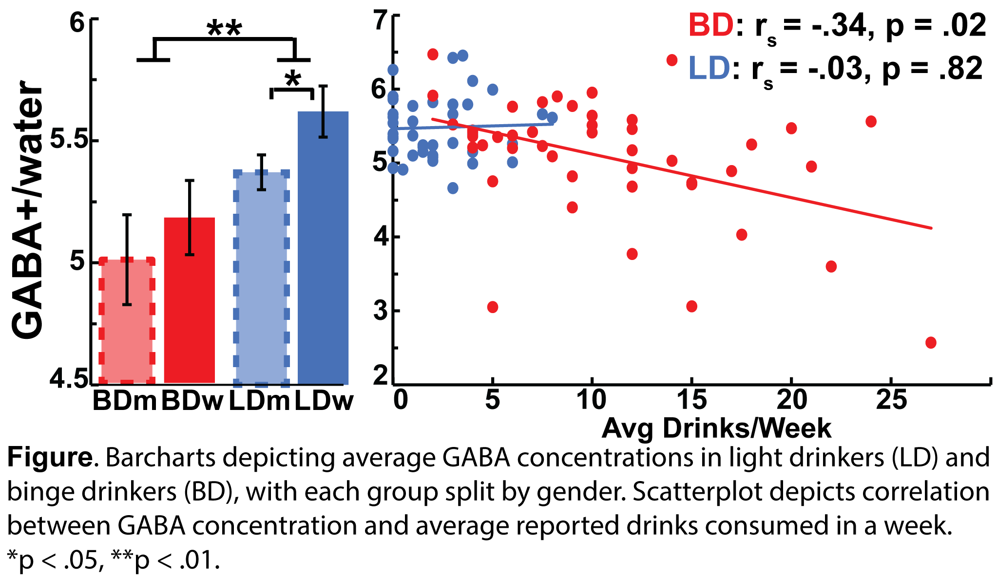
Barcharts depicting average GABA concentration in light drinkers (LD) and binge drinkgers (BD), with each group split by gender. Scatterplot depicts correlation between GABA concentration and average reported drinks consumed in a week.
GABA Concentration
Acute alcohol intoxication increases inhibition mediated by gamma-amino butryic acid (GABA) and decreases excitatory glutamatergic effects. It has been hypothesized that repeated and frequent binge drinking episodes leads to excitatory/inhibitory balance even while sober. A previous paper of from our lab found evidence of heightened excitability among sober binge drinkers, however we published another paper which found reduced GABA concentration directly using the MRS method. Additionally, it was found that GABA concentration correlated with average reported drinks per week.
Related Publications
Huang, S., White, D. R., & Marinkovic, K. (2022). Alterations of theta power and synchrony during encoding in young adult binge drinkers: Subsequent memory effects associated with retrieval after 48 h and 6 months. Front Psychol, 13, 1061016. doi:10.3389/fpsyg.2022.1061016. PDF
Marinkovic, K., Alderson Myers, A. B., Arienzo, D., Sereno, M. I., & Mason, G. F. (2022). Cortical GABA levels are reduced in young adult binge drinkers: Association with recent alcohol consumption and sex. NeuroImage: Clinical, 35, 103091. PDF
Correas, A., Cuesta, P., Rosen, B. Q., Maestu, F., Marinkovic, K.
"Compensatory neuroadaptation to binge drinking: Human
evidence for allostasis" Addiction Biology (Aug 2020) PDF
Holcomb L, Huang S, Cruz SM, Marinkovic K. (2019) Neural oscillatory dynamics of inhibitory control in young adult binge drinkers. Biological Psychology. PDF
Huang, S., Holcomb, L.A., Cruz, S.M., & Marinkovic, K. "Altered Oscillatory Brain Dynamics of Emotional Processing in Young Binge Drinkers." Cogn Affect Behav Neurosci (Nov 2017): 1-15 PDF
Molnar, S.M., Beaton, L.E., Happer, J.P., Holcomb, L.A., Huang, S., Arienzo, D., & Marinkovic, K. "Behavioral and Brain Activity Indices of Cognitive Control Deficits in Binge Drinkers." Brain Sci 8 no. 1 (Jan 2018) PDF
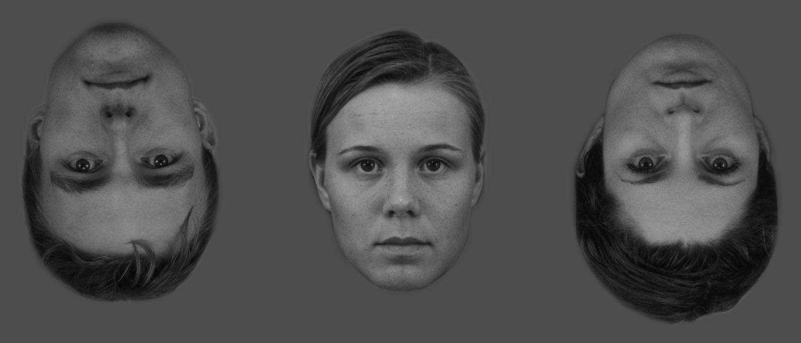
Face stimuli have captured a great deal of attention in the neuroimaging field, partly resulting from the hotly debated idea that faces are processed in a "face-specific" manner by dedicated brain areas. Indeed, efficient and correct appraisal of face identity and emotional expressions is of great importance to social primates, including humans. Our research and other evidence suggest that faces are processed in a series of successive stages engaging multiple brain areas in an interactive manner. The face-sensitive processing stage is activated at about 170ms after stimulus onset and is situated in the temporal lobe, between early visual cortex and later multimodal cortices that perform cognitive integration with the context.
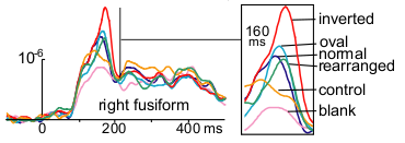
Group-based average time courses in response to a variety of stimuli. At ~160ms, inverted faces elicited the strongest activity in the right fusiform gyrus. Note that negative is up on the y-axis.
Related Publications
Marinkovic, Ksenija, Maureen G. Courtney, Thomas Witzel, Anders M. Dale, and Eric Halgren. "Spatio-Temporal Dynamics and Laterality Effects of Face Inversion, Feature Presence and Configuration, and Face Outline." Frontiers in Human Neuroscience 8:868 (Nov 10 2014).
Marinkovic, Ksenija, Marlene Oscar-Berman, Trinity Urban, Cara E. O'Reilly, Julie A. Howard, Kayle Sawyer, and Gordon J. Harris. "Alcoholism and Dampened Temporal Limbic Activation to Emotional Faces." Alcoholism-Clinical and Experimental Research 33, no. 11 (Nov 2009): 1880-92.
Halgren E, Raij T, Marinkovic K, Jousmäki V, Hari R. Cognitive response profile of the human fusiform face area as determined by MEG. Cereb Cortex. 2000; 10(1):69-81.
Klopp, J., K. Marinkovic, P. Chauvel, V. Nenov, and E. Halgren. "Early Widespread Cortical Distribution of Coherent Fusiform Face Selective Activity." Human Brain Mapping 11, no. 4 (Dec 2000): 286-93. PDF
Halgren, E., A. M. Dale, M. I. Sereno, R. B. H. Tootell, K. Marinkovic, and B. R. Rosen. "Location of Human Face-Selective Cortex with Respect to Retinotopic Areas." Human Brain Mapping 7, no. 1 (1999 1999): 29-37. PDF
Klopp, J., E. Halgren, K. Marinkovic, and V. Nenov. "Face-Selective Spectral Changes in the Human Fusiform Gyrus." Clinical Neurophysiology 110, no. 4 (Apr 1999): 676-82. PDF
 Language is essential to our communication with others and to our conceptualization of the world. Through words we acquire a multitude of information, articulate our thoughts or feelings and delight in mirth when sharing jokes. When comparing the neural dynamics of understanding written vs. spoken words, the activity starts in sensory-specific areas and progresses towards the simultaneously active supramodal regions in temporal and prefrontal brain regions. "Top-down" guidance from the prefrontal areas facilitates simultaneous lexical, semantic and contextual integration with the goal of rapid comprehension of verbal input.
The process of understanding a word in its current context peaks about 400 ms after word onset. It is carried out mainly through interactions of the temporal and inferior prefrontal areas on the left during word reading and bilateral temporo-prefrontal areas during speech processing. Neurophysiological evidence suggests that lexical access, semantic associations, and contextual integration may be simultaneous as the brain uses available information in a concurrent manner, with the final goal of rapidly comprehending verbal input. Because the same areas may participate in multiple stages of semantic or syntactic processing, it is crucial to consider both spatial and temporal aspects of their interactions to appreciate how the brain understands words.
Spatiotemporal pattern of reading a word
Spatiotemporal pattern of hearing a word
The two videos above are "brain movies" created from anatomically constrained estimates of MEG signal sources. These "brain movies" demonstrate the spatiotemporal pattern of activition when reading and hearing a word. The earliest activity represents the processing of basic sensory information. When reading a word, this activity starts at ~100ms in the occipital area. When hearing a word, activity starts earlier at ~55ms in the superior temporal region. Activity proceeds anteriorly via the ventral processing stream, and both modalities overlap in temporal and prefrontal regions. The peak of activity occurs at ~400ms, thought to signal accessing and integrating semantic representation into a contextual stream. Reading written words was left lateralized, while hearing spoken words engaged bilateral regions with left-dominant prefrontal activity.
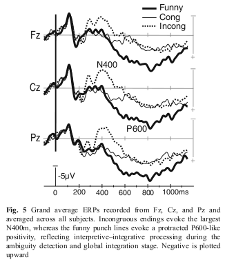
Grand average ERPs recorded from Fz, Cz, and Pz and averaged across all subjects. Incongruous endings evoke the largest N400m, reflecting interperetive-integrative processing during the ambiguity detection and global integration stage. Negative is plotted upward.
Language entails much more complexity than understanding individual words, as they are arranged in sentences and discourse. Understanding jokes, for example, relies on semantic, mnemonic, inferential, and emotional contributions of multiple brain areas. In addition to the left temporal and prefrontal regions, funny punch lines activate a "second take" processing in the right prefrontal cortex in order to detect and resolve the clever "twist" they contain. Coherent integration of the intended meaning and a sense of amusement may emerge from the dynamic interaction of these regions with special contributions from the right prefrontal region.
Related Publications
Marinkovic, Ksenija.. "Spatiotemporal Dynamics of Word Processing in the Human Cortex." Neuroscientist 10, no. 2 (Apr 2004): 142-52.
Marinkovic, Ksenija, Burke Q. Rosen, Brendan Cox, and Donald J. Hagler, Jr. "Spatio-Temporal Processing of Words and Nonwords: Hemispheric Laterality and Acute Alcohol Intoxication." Brain Research 1558 (Apr 16 2014): 18-32.
Marinkovic, Ksenija, Burke Q. Rosen, Brendan Cox, and Sanja Kovacevic. "Event-Related Theta Power During Lexical-Semantic Retrieval and Decision Conflict Is Modulated by Alcohol Intoxication: Anatomically Constrained Meg." Frontiers in Psychology 3 (2012 2012).
Marinkovic, Ksenija, Sharelle Baldwin, Maureen G. Courtney, Thomas Witzel, Anders M. Dale, and Eric Halgren. "Right Hemisphere Has the Last Laugh: Neural Dynamics of Joke Appreciation." Cognitive Affective & Behavioral Neuroscience 11, no. 1 (Mar 2011): 113-30.
Halgren, E., C. M. Wang, D. L. Schomer, S. Knake, K. Marinkovic, J. L. Wu, and I. Ulbert. "Processing Stages Underlying Word Recognition in the Anteroventral Temporal Lobe." Neuroimage 30, no. 4 (May 1 2006): 1401-13.
Marinkovic, K., R. P. Dhond, A. M. Dale, M. Glessner, V. Carr, and E. Halgren. "Spatiotemporal Dynamics of Modality-Specific and Supramodal Word Processing." Neuron 38, no. 3 (May 8 2003): 487-97.
Dhond, R. P., R. L. Buckner, A. M. Dale, K. Marinkovic, and E. Halgren. "Spatiotemporal Maps of Brain Activity Underlying Word Generation and Their Modification During Repetition Priming." Journal of Neuroscience 21, no. 10 (May 15 2001): 3564-71. PDF
Our work on Autism Spectrum Disorder (ASD) uses the combined spatial and temporal strengths of anatomically constrained MEG (aMEG) to assess neural functioning. This on-going series of publications utilizes several tasks administered during scans which probe language processing and cognitive functions.
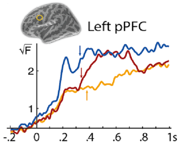
Timecourses depict averaged activity to all words for typically developing children (TD), and kids with low (l-ASD) and high (H-ASD) functioning ASD. Arrows indicate onset of N400m. Yellow circle shows estimate of MEG signal, in left prefrontal cortex. N400m are delayed to all children with ASD, and H-ASD had diminished N400m amplitude.
Language
ASD is characterized by difficulties in social communication, and language impairments are common. A sample of adolescents with ASD and typically developing peers took part in a lexical decision task. The task involved the presentation of standard words (SW), animal words (AN), and pseudo words (PW, e.g. "stigor"). Adolescents with ASD demonstrated bilateral activations, possibly as a compensation mechanism. Additionally, there was a diminished N400 (marker of linguistic processing) in children with ASD in the left LTC. Furthermore, the N400s of lower-performing children with ASD were less differntiated between conditions, and their N400 latency was delayed. This all points to impaired lexico-semantic processing.
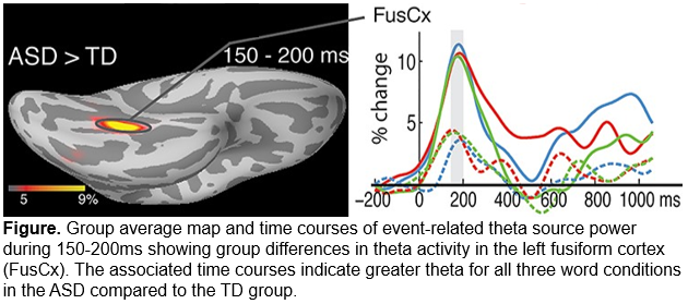
Group average map and time courses of event-related theta source power during 150-200ms showing group differences in theta activity in the left fusiform cortex (FusCx). The associated time courses indicate greater theta for all three word conditions in the ASD compared to the TD group.
Response Conflict
The lexical decision task mentioned above also required participants to respond to standard words (SW) and animal words (AN) using their left index and middle fingers respectively. Adolescents with ASD exhibited greater theta activity in left temporal and frontal cortical areas and the anterior cingulate cortex. These findings suggect compensatory recruitment to offset inefficient lexico-semantic retrieval under cognitively demanding conditions, as well as suboptimally organized motor circuitry.
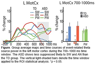
Group average time courses of event-related theta source power in the left motor cortex in the left motor cortex (L MotCx). Barcharts depict average theta within the 700-1000ms time window. The ASD group shows less suppressed theta to SW and AN than the TD group. *p<.05.
Related Publications
You, Y., Correas, A., White, D. R., Wagner, L. C., Jao Keehn, R. J., Rosen, B. Q., . . . Marinkovic, K. (2023). Mapping access to meaning in adolescents with autism: Atypical lateralization and spatiotemporal patterns as a function of language ability. NeuroImage: Clinical, 39, 103467.
Yuqi You, Angeles Correas, R Joanne Jao Keehn, Laura C Wagner, Burke Q Rosen, Lauren E Beaton, Yangfeifei Gao, William T Brocklehurst, Inna Fishman, Ralph-Axel Müller, Ksenija Marinkovic, MEG Theta during Lexico-Semantic and Executive Processing Is Altered in High-Functioning Adolescents with Autism, Cerebral Cortex, (Oct 19 2020). https://doi.org/10.1093/cercor/bhaa279
In conjunction with Dr. Thereasa Cronan's lab, we used electroencephalography (EEG) to investigate the underlying causes of fibromyalgia syndrome (FMS). FMS is associated with chronic pain and cognitive problems often known as "fibro fog". Because analgesics have been shown to be ineffective at treating the pain associated with FMS, it has been proposed that the pain-related symptoms are instead related to central nervous system dysregulation.
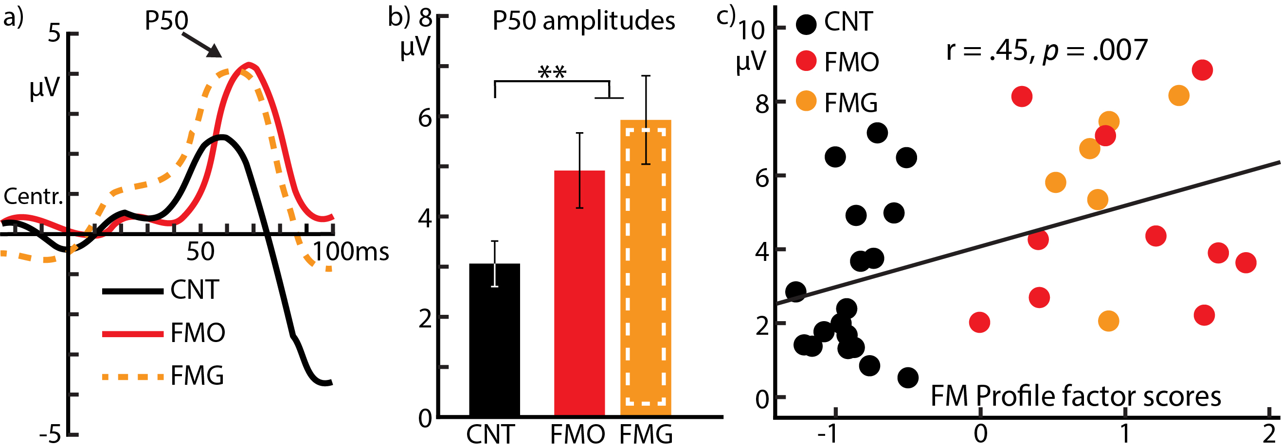
ERPs: Somatosensory domain. a) Somatosensory ERPs to the sensation averaged over central electrodes for all three groups. The P50 deflection is marked by an arrow. Negative is down on the y-axis. b) Histograms display mean +/- SEM of P50 amplitudes. c) Scatter plot shows correlation between P50 amplitudes and the FM profile PCA factor. People with FM had greater P50 amplitudes than other healthy matched adults. **p<.01
Acute Hypersensitivity
A sample of mostly women with FM, as well as a matched non-FM control group, took part in a task which combined auditory and somatosensory stimuli, designed as a classical conditioning experiment. Participants first heard either a high or low tone, and were administered an "uncomfortable, not painful" stimulation to their shin, depending on the tone pairing. Participants with FM were more sensitive to electrical sensations and audio tones. Additionally, they also had more extreme P50 and N100 amplitudes as well as greater electodermal reactivity. A subset of FM participants who took GABA agonist medication had less extreme N100s and reduced electrodermal reactivity. This suggests basic hyperexcitability in people in FM, and some commonly used medications for FM treatment may work by normalizing this excitability.
Related Publications
Marinkovic, K., Woodruff, D., White, D. R., Caudle, M. M., & Cronan, T. (2023). Neural indices of multimodal sensory and autonomic hyperexcitability in fibromyalgia. Neurobiology of Pain, 14, 100140.
During the COVID-19 pandemic, our lab started a project in order to investigate the neural indices of long COVID, also referred to as post-acute sequelae of COVID-19 (PASC). This is an on-going project spanning multiple technologies, methods and tasks. The sample included in the results below was composed of by mostly, otherwise healthy adults who reported mild PASC symptoms after clearing acute COVID-19 infection. The results also involve the use of previously acquired control groups.
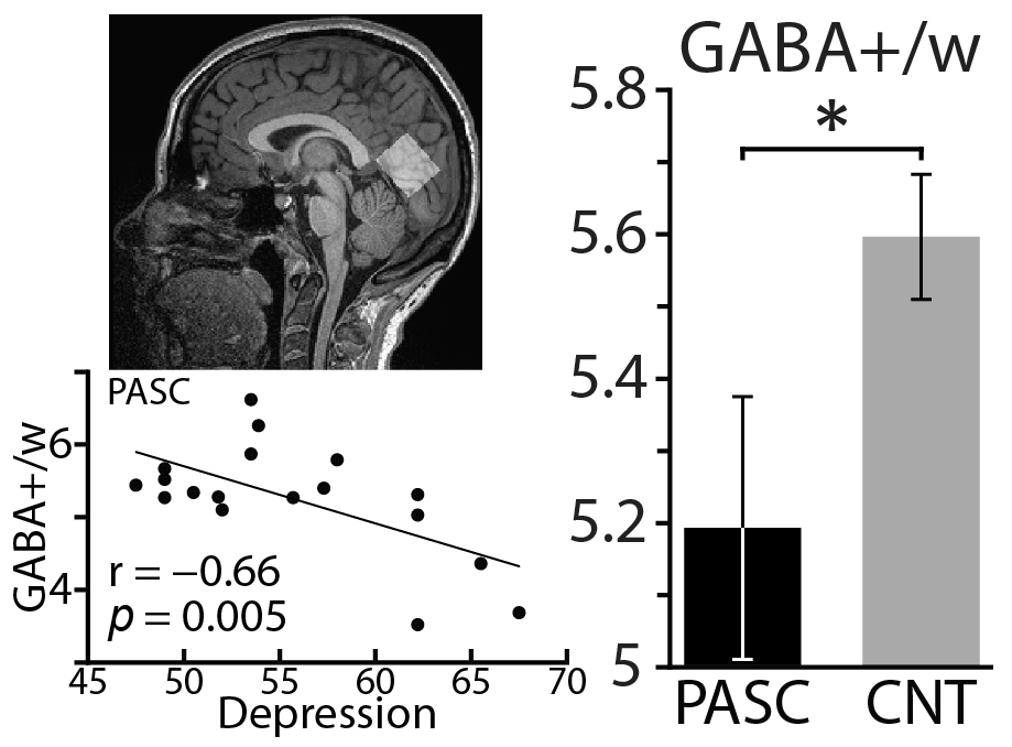
Barchart depicting average GABA referenced to water in the PASC and control groups. Scatter plot depicts correlation between GABA concentration and depression. Source of GABA measurement in the brain is shown with white rectangle.
Reduced GABA
Participants recieved an MRS scan to determine the concentration of GABA related metabolites in vivo. GABA is the most common neurotransmitter and is ciritcal for regulation of neural activityIt was found that participants with PASC had less GABA than the control sample, and measures of GABA negatively correlated with depression. Lowered GABA suggests cortical hyperexcitability, which may result from neuroinflammation stemming from the body's immune system response to COVID-19. This hyperexcitability may underlie common PASC symtpoms, such as depression.
Related Publications
Marinkovic, K., White, D. R., Alderson Myers, A., Parker, K. S., Arienzo, D., & Mason, G. F. (2023). Cortical GABA Levels Are Reduced in Post-Acute COVID-19 Syndrome. Brain Sciences, 13(12), 1666.
We have investigated differences in dopamine availability, referenced by participants' genotype for the COMT gene. This functional polymorphism may modulate the neural circuitry underlying inhibitory control, thus giving rise to heritable differences in behaviors. This gene codes for the amount of COMT enzyme is produced, which breaks down dopamine in the synapse. In a study utilzing anatomically constrained MEG and a Go/NoGo response inhibition task, we compared differences in neural function between participants Met/Met (high dopamine) and Val/Val (low dopamine).
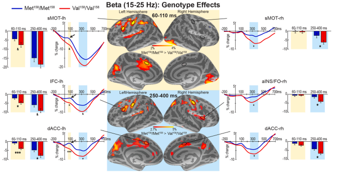
Histograms depict average beta power in various brain regions involved in motor preparation and planning during an early (60-110ms) and a later (250-400ms) window, averaged across all trials. Timecourses show the average change in beta change from baseline, and brain maps show source of signals. Val/Val consistently showed greater beta reduction than Met/Met, indicative of greater impulsivity.
Beta oscillations usually decrease prior to making a response, and are thought to reflect engagement of the motor cortices, basal ganglia, and other areas contributing to motor planning, inhibition, and execution. Val/Vals showed generally greater beta decrease, and may indicate greater preparedness to respond and impulsivity. This greater decrease correlated with extraversion and greater drinking. In addition, higher beta in Met/Mets may be the result of a "pause" before response selection, particarly when looking at Go trials with slow reaction times, which was absent in errorneous NoGo responses.
Related Publications
Happer, J. P., Beaton, L. E., Wagner, L. C., Hodgkinson, C. A., Goldman, D., & Marinkovic, K. (2024). Neural indices of heritable impulsivity: Impact of the COMT Val158Met polymorphism on frontal beta power during early motor preparation. Biol Psychol, 191, 108826.